
Electron microscopy (EM) has transformed our ability to visualise the microscopic world, revealing the intricate architecture of cells, pathogens and biomolecules at extraordinary resolution. The revolution in cryogenic electron microscopy (cryo-EM), recognised by the 2017 Nobel Prize in Chemistry, has fundamentally changed structural biology by enabling scientists to determine the three-dimensional structures of biological molecules with unprecedented detail. During the COVID-19 pandemic, cryo-EM played a pivotal role in revealing the structure of the SARS-CoV-2 spike protein, accelerating vaccine development and structure-based drug discovery.
However, although South Africa has a strong tradition in EM, researchers still rely on overseas facilities for high-resolution studies, limiting scientific competitiveness, advanced training, and rapid responses to emerging health challenges.
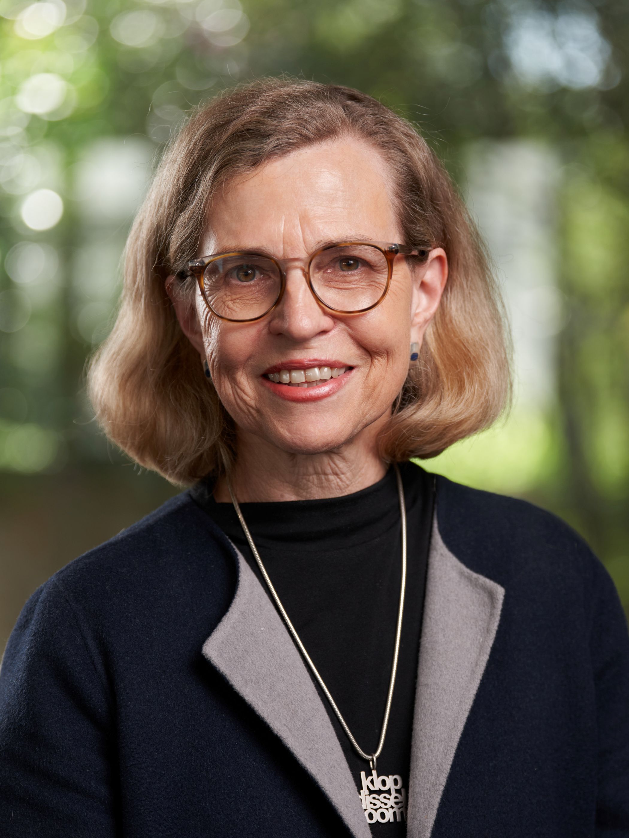
In the first public lecture of the semester, Lydia-Marie Joubert made a strong, convincing argument for the urgent establishment of a national cryo-EM platform in South Africa, which she described as an investment in scientific infrastructure, education, healthcare, pharmaceutical innovation and human capacity development, positioning the country as a regional leader in structural biology and biomedical research, and strengthening Africa's role in the global scientific community.
Joubert is Director of Operations at the Stanford-SLAC Cryo-Electron Tomography Center, where she leads operations, training and technology development in cryo-electron tomography and advanced cryo-EM. She is also Professor Extraordinary in Microbiology at Stellenbosch University. She obtained her BSc in Mathematics and Botany, Honours BSc, and MSc in Botany from Stellenbosch University, followed by a PhD jointly supervised by the University of Pretoria and Indiana University. During her MSc, she completed a research internship in Plant Genetics at the Weizmann Institute of Science in Israel. Later in her career, she also obtained an MPhil in Higher Education.
Her research focuses on developing and applying cutting-edge microscopy approaches to answer fundamental biological questions across plant, microbial and biomedical sciences. Her work advances cryo-electron tomography, cryo-focused ion beam scanning EM, correlative light and EM, and three-dimensional imaging workflows that enable visualisation of biological structures at sub-nanometre resolution. She has contributed to research in plant development, environmental microbiology, biomaterials, bioenergy and cell biology in health and disease. She is widely recognised for excellence in scientific visualisation, with numerous international awards for microscopy imaging, and her electron micrographs have appeared on the covers of leading scientific journals.
“Cryo-EM has transformed what is possible, but access remains deeply uneven globally,” she said. “It’s about scientific independence, being able to address important questions and participate as an equal partner in global science. It represents far more than the acquisition of a sophisticated microscope. It is an investment not only in our scientific future, but in South Africa’s future.”
The history of cryo-EM imaging dates from 1968, when De Rosier and Klug published the first 3D reconstruction from electron micrographs; followed in 1975 by Frank’s foundational image processing methods, which made cryo-EM more widely usable, and Henderson, who achieved atomic resolution. In the early 1980s, Jacques Dubochet developed vitrification–fast freezing of samples without the formation of ice crystals; the 1990s – 2000s were called the ‘blobbology era’, when most structures could be resolved to ‘blobs’ of only 20 to 30 Å; while in 2012 and 2013, direct electron detectors triggered the resolution revolution, pushing resolution past 4 Å. In 2017, the Nobel Prize in Chemistry was awarded to Dubochet, Frank and Henderson for developing cryo-EM. Today, computational reconstruction and atomic modelling mean that 2 to 4 Å structures are routine and Cryo-EM Protein Data Bank entries have grown 85-fold.
Joubert explained that resolution is the smallest distance between two objects that allows you to distinguish them as separate entities. It’s defined by the wavelength of the light source. “Since the wavelength of an electron is much shorter than that of visible light, you can achieve much higher resolution with electron microscopy.”
Cryo-EM measures at the level of angstroms (Å, named after Swedish physicist Anders Jonas Ångström) − a measure of a 100 millionth of a centimetre. This means we can understand in detail, via high resolution, how viruses and parasites infect cells.
“With viruses, for example, you can see the movement and bending angles of spike proteins and therefore understand their infectivity, virus fitness and visualise antibody binding,” explained Joubert.
“It’s vital to vaccine development – by seeing the enemy and knowing how to target it better,” explained Joubert. “Currently, Africa imports 99% of vaccines in use. We also need more vaccines because of the predominantly youthful population on the continent. This makes it hard to prepare healthcare systems for outbreaks. We can’t rely on global supply chains. We need national strategies to guarantee health security in Africa, and cryo-EM should be part of these.”
Cryo-EM gives us a more complete picture of life, and this extends to beyond human life. “You can also do things like visualise photosynthetic complexes – such as those found in plants and green algae. You can view at the atomic level what happens in the photosynthetic cell and see the effect of different environmental conditions on the processes underlying photosynthesis,” said Joubert.
The latest Cryo-EM instrumentation has a workflow ranging from sample preparation to data collection, to image processing and model building, refinement and validation, resulting in a 3D, high-resolution structure for biological interpretation.
Africa still a spectator
“Cryo-EM plays an essential role in drug and vaccine development, shortening the path to developing therapeutics and reducing the disease burden,” said Joubert. But she pointed to the frustration of knowing the technology is available and can impact health but is not available for everyone – “There is only one Cryo-EM instrument available on the continent, and that is at the tip of Africa – at the University of Cape Town - and it’s not an advanced state-of-the-art instrument – still requiring part of the workflow to be done in the UK.”
Even though South Africa has a long history of EM dating back to the 1940s − Aaron Klug, who was raised and educated in South Africa, participated in the first 3D reconstruction and won the 1982 Nobel Prize for Chemistry for his development of crystallographic EM – the country has fallen behind in the race to advanced cryo-EM. “There are multiple EMs in South Africa, but only one (ageing) cryo-TEM,” said Joubert. “We need the most advanced instruments and to broaden access.”
This means there is only one cryo-EM facility on the continent serving 54 countries; turnaround times are slow due to sample shipping costs as well as delay and degradation risks, all resulting in too few local training pipelines for structural biologists. Joubert pointed out that the latest machines cost approximately US$ 5 million, so it’s not an unreasonable investment but, of course, also requires infrastructure, dedicated staff and ongoing running costs.
“The global community will only reach out after an initial government investment,” she continued. “We need local infrastructure and funding, capacity development and retention and access to international infrastructure. South Africa needs a roadmap, policy-making and long-term sustainable funding.”
She also pointed to the urgent need to retain capacity and expertise in the country and on the continent. “There has been an over 205% increase in black STEM graduates since 2005, but there is still the massive problem of a brain drain because these graduates can’t do the work here.”
“If you have the technology, people come for training and can do research ‘at home’ in South Africa,” she added. “We need to inspire and inform the next generation, make this work attractive to young people, show that we are not isolated but part of international networks. Now is a good time to invest in cryo-EM and join the global community because the early mistakes in the field have already been made – the field has matured, and many challenges are now well understood. Cryo-EM is not merely an interesting specialisation but a source of solutions.
Cryo-EM can play a huge role in multiple African health priorities, including endemic infections, antimicrobial resistance, emerging and epidemic viruses (including TB, HIV/AIDS and Malaria), non-communicable disease, one health and veterinary programmes, biotechnology and neglected diseases
“A single microscope can support research spanning infectious disease, cancers, metabolic and neurological disease rather than being tied to a single disorder,” said Joubert. “We need to step up and do it ourselves. We have the potential to change the world.”
Article: Michelle Galloway
Picture: Curt Ruiters, Quickclick Productions